sábado, 19 de maio de 2018

Historical Reading: The California Gold Rush


Historical Reading: The California Gold Rush

TwitterPrintEmailPinterestMore

Placeholder Alt Text
This illustration depicts life in a miner’s camp along the foothills of the Sierra Nevada mountains. From Ballou’s Pictorial Drawing-Room Companion (1856).
Gold was discovered in California by James Marshall at Sutter’s sawmill on the South Fork of the American River near Coloma (36 miles northeast of Sacramento) on Jan. 24, 1848. The first published accounts of the find appeared in “The Californian,” a San Francisco newspapers, on March 15, 1848. The news was first met with disbelief by those who doubted this valuable metal could just be picked up off the ground.
Subsequent confirmation of the initial reports of the extent of the gold region set off a rush.
Adventurers from the U.S. and around the world traveled to California to seek their fortunes. Excitement at their financial prospects was compounded by a desire to get there as quickly as possible. They borrowed money, mortgaged their property, and spent their life savings to make the arduous journey. Some made their fortunes, but many did not.
Most began their journey in the Eastern U.S. and departed via three routes:
  • By ship to Central America, followed by a land crossing at the Isthmus of Panama, and then another ship to San Francisco (three to five months in 1850).
  • By ship around Cape Horn in South America and on to San Francisco (five to eight months).
  • By travel westward across the plains from the U.S. or via Mexico (three to four months).
This arrival of thousands of “prospectors” transformed and accelerated the development of the territory of California (including its admission as the 31st state in 1850) within a few years. Mining camps and towns sprang up throughout the interior region, with towns of Sacramento and Stockton as the gateways to the mining areas. Numerous reports on the occurrence and mining of gold along the western side of the Sierra Nevada Mountains were written, and a number of participants later published accounts of their mining exploits.
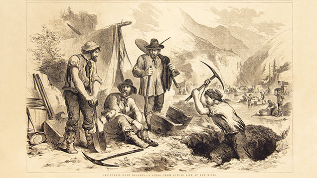
Gold seekers mined by panning in the rivers and streams, using the flowing water through an oblong box that was rocked back and forth, to carry off the lighter sediments. High-pressure hydraulic hoses were later used to wash gold from hillsides. Eventually, dredging of the larger rivers was undertaken, and underground mines were dug to reach the gold ore. Gold mining in California reached its peak production in 1852, and gradually declined thereafter.

HOW TO USE THIS READING LIST

This reading list was compiled to give you an opportunity to learn more about the history of the California Gold Rush. A number of the articles were published in the 1800s and early 1900s – when many classical gem deposits of historical importance were discovered – and gemology and mineralogy became sciences. The list is presented in chronological order to emphasize the development of ideas over time. The list is not comprehensive, but a compilation of the some interesting gemological information that has often been forgotten or overlooked.
Many of the articles exist in the public domain and can be found online at digital libraries such as Hathitrust, Internet Archive, or other digital repositories. More recent publications can often be found in libraries, including the Richard T. Liddicoat Gemological Library. Abstracts of these articles can usually be found on the website of the original journal or magazine, and the article itself is often available for purchase from the publisher.
Regarding the GIA library’s holdings and on-site access, please contact the GIA library in Carlsbad.
Gold, Gold, Author unknown, Scientific American, Vol. 4, No. 1, p. 2, (1848). An early report about six months after the event “of the discovery of an immense bed of gold one hundred miles in extent, on the American Fork and Feather rivers”.
The Golden Land, Author unknown, Scientific American, Vol. 4, No. 13, p. 98, (1848). “A short time ago, the most flattering accounts were received in this city from California about the mountains of gold and the valleys flowing with silver. Some believed it was a joke, while others believed it to be a ‘hue and cry’ for some speculative purpose, and to the latter implication we must plead guilty. We believed that the accounts received here a short time ago about vessels being deserted by their crews and houses by their inhabitants, who had proceeded to the El Dorado valley, were all a hoax or something worse. But it seems, after all, that Madam Rumor sometimes tells true tales. The golden hills of California it seems are not imaginary elevations, but bona fide treasure houses.”
Gold and Gold Washings, Author unknown, Scientific American, Vol. 4, No. 15, p. 114, (1848). “The gold region of California is said to extend on both sides of the Sierra Nevada as far south as the headwaters of the San Joaquin River – a distance of 400 miles in length and 100 in breadth.” A short description of how the gold is found and mined is provided.
California Gold, Author unknown, Scientific American, Vol. 4, No. 18, p. 141, (1849). “The gold excitement is as strong in our city as ever. In one day last week ten vessels sailed from this port.” The report mentions many different types of people are traveling to the gold fields. “It is calculated that no less than 150,000 emigrants will be on their way to California from the States in two months.”
Gold-Finding in California, Author unknown, Chambers’s Edinburgh Journal, Vol. 11, No. 265, pp. 61-62, (1849). This is one of the first descriptions of the California gold rush published in Europe. Following the discovery in the spring of 1848, the area east of Sacramento became “a scene of busy gold finding, for which perhaps no parallel exists in the history of any country. One is at first tempted to suppose the whole affair a popular delusion, or a deliberate exaggeration, after a well-known transatlantic manner, but such theories are no longer tenable … As soon as it was known that gold was literally to be had for the lifting of certain parts of the country, an almost universal abandonment of the common pursuits of life took place.”
“It will remain to be seen whether this extraordinary windfall will prove of any serious permanent benefit to America or any of her citizens. History has shown that gold-finding has never yet been a permanently advantageous pursuit. If America thrives by picking up this precious metal in the wilds of California, she will be an exception from a pretty well-established rule.”
America, Author unknown, Gentleman’s Magazine, Vol. 186, (February), p. 192, (1849). A brief report about the gold rush, which begins as, “The new world, and we may add the old also, has been thrown into a whirl of excitement by the abundant discovery of surface gold on the plains of Upper California.”
The Gold-Washings of California, Author unknown, New Monthly Magazine, Vol. 85, No. 338, pp. 252-254, (1849). A discussion of a report of Edwin Bryant, a resident of San Francisco, about the future settlement of California by those who were coming to prospect for gold.
Account of the Gold Region, Author unknown, Littell’s Living Age, Vol. 20, No. 248, pp. 305-308, (1849). The account of a newspaper reporter from New Orleans who visited San Francisco and then the gold diggings around Sutter’s Mill in the summer of 1848.
The Apoplexy of Gold, Author unknown, Littell’s Living Age, Vol. 20, No. 249, p. 371, (1849). A discussion of the frenzied excitement brought on by the discovery of gold.
California Fever in England, Author unknown, Littell’s Living Age, Vol. 20, No. 249, p. 371-372, (1849). A discussion of advertisements in British newspapers for the arrangements of ships to transport fortune-seekers to California.

Fonte:Gems & Gemology

Iridescence in Metamorphic “Rainbow” Hematite

Iridescence in Metamorphic “Rainbow” Hematite


Rainbow hematite from Brazil’s Andrade mine.
Figure 1. Rainbow hematite from the Andrade mine in João Monlevade, Minas Gerais, Brazil. Photo © Rock Currier, Mindat.org.
The authors investigated “rainbow” hematite from Minas Gerais, Brazil, using electron microscopy, atomic force microscopy, and synchrotron X-ray diffraction to determine the cause of its intense wide-angle iridescence. The study revealed that the interference is produced by a highly periodic microstructure consisting of spindle-shaped hematite nanocrystals containing minor Al and P impurities. The nanorods are 200–300 nm in length and 50–60 nm in width. They are arranged in three orientations at 120º angles with respect to each other and stacked layer by layer to form the bulk crystal. The distances between adjacent parallel spindle-shaped particles within the same layer fall in the range of 280–400 nm, generating a diffraction grating for visible light. The organized substructure is apparent on all freshly fractured surfaces, suggesting that it represents more than an exterior surface coating. The authors propose that this periodic substructure results from arrested crystal growth by the oriented aggregation of hematite nanorods.
Hematite specimens that frequently display iridescence are described as “rainbow hematite” and “turgite” (figure 1). The latter term originated with the German mineralogist Rudolph Hermann, who coined it in 1844 to describe iron hydroxide specimens found near the Turginsk River in the Ural Mountains (Hermann, 1844). “Turgite” was discredited as a distinct mineral name in the 1920s, however, based on thermogravimetric (Posnjak and Merwin, 1919) and X-ray diffraction (Böhm, 1928; Palache et al., 1944) studies that identified such specimens as mixtures of microcrystalline hematite (Fe2O3) and either goethite (FeOOH) or amorphous Fe hydroxide. Nevertheless, “turgite” has been retained by the mineral collecting and gem community as a catch-all term for naturally iridescent iron (hydr)oxide minerals.
Rock Currier and colleague at the Andrade mine.
Figure 2. Rock Currier (left) and colleague survey a seam of rainbow hematite at the Andrade mine. Photo © Rock Currier, Mindat.org.
Mineral dealer Rock Currier (1940–2015) was largely responsible for introducing rainbow hematite to the U.S. market. According to notes he published on Mindat.org, Currier first visited an outcrop of what he called “color rock” from the Andrade mine in João Monlevade, Minas Gerais, Brazil, in 1991 (figure 2; Currier, 2012). Shortly after, he shipped a truckload of 55-gallon barrels filled with an estimated 15 tons of the material to the United States. Initially he attempted to sell the rainbow hematite “by the barrel for $3 per pound without very much luck.” At a subsequent Tucson Gem and Mineral Show, however, he “arrived to find some guy had rented a big billboard and was selling the stuff as the latest and greatest metaphysical jewelry item. One guy was backing little pieces of the stuff with obsidian and selling earrings…for $90 a pair.” Fortunately, Currier had kept enough of the rainbow hematite to select higher-grade samples, and he saw brisk sales of individual pieces.

Bulk samples of this Brazilian rainbow hematite are still sold at major mineral and gem shows, but our discussions with dealers indicate a growing scarcity. Currier attempted to purchase more material from the mine, but apparently the major seam of rainbow hematite at Andrade underlies the primary mine haul road, and excavation would have destabilized this conduit to the open pit. Nevertheless, exquisite pieces of rainbow hematite jewelry are still sold online, with accent stones such as amethyst, apatite, sapphire, tourmaline, and tsavorite (figure 3). Moreover, rainbow hematite plays an important role in the local culture. Villages neighboring the city of Belo Horizonte traditionally sprinkle the streets with truckloads of powdered material for festivals, creating an effect that Currier (2012) likened to “standing in a pile of peacock feathers.“
Rainbow hematite in pendant (left) and ring (right).
Figure 3. Left: An 18K gold pendant featuring rainbow hematite from Minas Gerais, Brazil, accented with amethyst and apatite. Right: Rainbow Rapture ring in 18K gold with tourmaline, sapphire, and tsavorite. Designs by Judith Anderson. Photos by The Jewelry Experts, Bijoux Extraordinaire, Ltd.
To our knowledge, Ma and Rossman (2003a,b) have performed the only scientific investigation into the cause of iridescence in rainbow hematite, analyzing specimens from Brazil, Mexico, Italy, and several sites in the United States. Their results are also briefly highlighted in Nadin (2007). Using field-emission scanning electron microscopy (FESEM), Ma and Rossman (2003b) reported that their rainbow hematite specimens were coated with a “thin film” of rod-shaped nanocrystals, each measuring 5 to 35 nm in thickness and hundreds of nm in length. These nanocrystals were oriented in three directions, at 120º angles with respect to each other, and formed a grid-like network. The nanocrystals were too small to determine precise chemical compositions, but energy-dispersive spectroscopy (EDS) revealed high concentrations of Al and P in a ratio that varied from 2.2 to 3.8. Ma and Rossman (2003b) noted that “the minute crystals have failed to produce either an X-ray powder diffraction pattern, an electron back-scatter diffraction pattern, or a Raman spectrum.” The study interpreted the rod-like nanocrystals as a new mineral, but a full characterization was not possible with the resolution of the instrumentation.

Over the 15 years since these results were published online, more sophisticated analytical techniques have become available. For the present study, we sought to determine whether iridescence in rainbow hematite arises from thin-film effects involving Al phosphate phases, as earlier researchers hypothesized, or whether a different mechanism is involved. Rainbow effects in minerals are commonly attributed to nanoscale coatings, as in bornite (Buckley and Woods, 1983; Vaughan et al., 1987) and fire obsidian (Ma et al., 2001, 2007). Yet many gem materials (e.g., opal, labradorite, iris agate, and iris quartz) contain modular substructures that create a diffraction grating for visible light and generate rainbow effects (Darragh et al., 1966; Miura and Tomisako, 1978; Heaney and Davis, 1995; Lin and Heaney, 2017). These substructures often yield insights into exotic crystal growth processes that can inspire pathways for the self-assembly of synthetic materials. For the present investigation, we examined rainbow hematite from the Andrade mine using a combination of light optical microscopy, X-ray diffraction (XRD) and Rietveld analysis, FESEM, and atomic force microscopy (AFM).
Specimen Description. We purchased the iridescent hematite samples used in this study at the 2014 Tucson Gem and Mineral Show from Cinderhill Gems (Meadow Vista, California), who traced the material to Rock Currier’s bulk shipment from Brazil in the 1990s. The specimens we studied appeared identical in macroscopic physical character to those described by Currier (2012). As noted below, the microstructures matched the descriptions for Andrade hematite in Rosière et al. (2001) and Ma and Rossman (2003b). Thus, we concluded that the rainbow hematite in this study indeed derives from the Andrade mine.
Geologic map of the Quadrilátero Ferrífero.
Figure 4. (A) Geologic map of the Quadrilátero Ferrífero in the southern portion of the São Francisco Craton (SFC), Brazil. Iridescent hematite derives from the Andrade mine, denoted toward the upper right as MAN. WLSD: Western Low-Strain Domain; EHSD: Eastern High-Strain Domain. IS: Itabira Syncline. From Mendes et al. (2017), as modified from Dorr (1969).
The Andrade iron ore deposit is located in the eastern high-strain domain of the Quadrilátero Ferrífero district (figure 4) in the southern part of the São Francisco Craton of Brazil (de Almeida, 1977). The Quadrilátero Ferrífero (or “Iron Quadrangle”) is so called because the Paleoproterozoic metasediments in the Minas Supergroup exhibit a rectangular arrangement of synclines (Rosière et al., 2001; Rosière and Chemale, 2008). The Caraça, Itabira, Piracicaba, and Sabará groups are four sequences of the Minas Supergroup rocks (Dorr, 1969; Mendes et al., 2017; see figure 5). The iron ore deposits are located within metamorphosed banded-iron formations in the Cauê Formation of the Itabira Group; the Andrade mine is in a contact-metamorphic zone within this formation. The rainbow hematite occurs as iridescent, specular seams oriented parallel to bedding. The material is brittle and fractures into lath-like splinters, but the crystals within the laths have a granoblastic texture, a term used to describe equigranular minerals without sharp crystal faces in metamorphic rocks. Rosière et al. (2001) attribute these textures to post-tectonic deformation and recrystallization.
Stratigraphic column of the Quadrilátero Ferrífero.
Figure 5. Stratigraphic column of the Quadrilátero Ferrífero. Rainbow hematite is found within the Cauê banded-iron formation. From Carlos et al. (2014).
The Andrade rainbow hematite seam occurs within a banded-iron formation (BIF) in the Itabira Group (again, see figure 5), which has lent its name to a broad class of metamorphic rocks called itabirites. These are hematitic (rather than mica) schists that formed when the original jasper bands in the BIF recrystallized into distinct layers of macroscopic quartz and hematite (or sometimes magnetite). The hematite schists, from which we infer the rainbow hematite derives, are intergrown within itabirites (Barbour, 1973).

Scanning Electron Microscopy and Energy-Dispersive Spectroscopy. SEM and EDS were used to characterize surface topography and compositional information for the specimen. As iridescence was evident even from freshly fractured surfaces of the Andrade specimen, we removed a flake from one of our specimens and affixed the flake to an SEM mount using carbon fiber tape so that the flat iridescent surface was parallel to the surface of the SEM mount. We used an FEI Nova NanoSEM 600 field emission scanning electron microscope, outfitted with an Oxford Instruments X-Max (Model 51-XMX1105) silicon drift detector used for EDS analysis, in the Materials Characterization Laboratory (MCL) at Pennsylvania State University to examine the iridescent hematite. Nanoscale secondary electron images were obtained at an acceleration voltage of 5 kV and a current of 9 pA. EDS data were processed using the Oxford Instruments NanoAnalysis AZtec software (version 2.4). We chose three different accelerating voltages (20, 10, and 5 kV) to acquire spectra for the same sites.

Atomic Force Microscopy. To obtain high-resolution surface topography, we used a Bruker Icon I AFM (MCL, Pennsylvania State University) in PeakForce Tapping (PFT) mode with the ScanAsyst image optimization technique. In PFT mode, the cantilever is brought in and out of contact with the surface. PFT algorithms can precisely control the instantaneous force interaction, allowing the use of reduced forces in the imaging process. In this way, both fragile probes and samples can be protected without compromising image resolution. ScanAsyst automatically adjusts the appropriate parameters (such as set points, feedback grains, and scan rates) during the scan and continuously monitors image quality. The AFM probe for this analysis was a Bruker ScanAsyst-Air probe, which has a silicon tip on a nitride lever. The front angle of the tip was 15° and the back angle was 25°. For AFM imaging, the peak force set point ranged from 1 to 4 nN and the scan rate was 1 to 0.5 Hz, with 512 data points per line in each scan. NanoScope Analysis software (version 1.50) was used to process the AFM data.

X-Ray Diffraction. Hematite was powdered in an agate mortar and pestle under acetone. Upon drying, the powder was loaded into a 0.7 ID polyimide capillary for X-ray diffraction. Synchrotron X-ray diffraction data were collected at the GeoSoilEnviroCARS (GSECARS) 13-BM-C beamline at the Advanced Photon Source (APS), Argonne National Laboratory (Argonne, Illinois). The X-ray wavelength was 0.8315(4) Å, and the detector-sample distance was 100.469(1) mm. XRD data were collected with a MAR165 CCD camera. The diffraction pattern was integrated into intensity vs. 2θ plots using the Fit2D program with a polarization factor of 0.99 (Hammersley et al., 1996).

Structure Refinement. Rietveld refinement is a technique for determining atomic structures by comparing the misfit between an observed powder X-ray diffraction pattern and a diffraction pattern calculated on the basis of a model crystal structure (Rietveld, 1969). Factors such as unit-cell parameters and atom positions are allowed to refine until the misfit between the observed and calculated patterns is minimized. Because we suspected that the atomic structure of the iridescent hematite from the Andrade mine might deviate from ideal hematite, we applied Rietveld analysis of the synchrotron XRD data to refine the crystal structure. The Rietveld software employed was the EXPGUI interface (Toby, 2001) of the general structure analysis system (GSAS) (Larson and Von Dreele, 1994). To obtain instrumental broadening parameters, we refined the structure of a LaB6 standard using starting parameters from Ning and Flemming (2005). For the hematite refinement, we incorporated the peak profile parameters refined for LaB6, and the initial structure parameters for hematite came from Blake et al. (1966). Using a 2θ range of 11.5° to 37.5° (dhkl = 4.1 Å to 1.3 Å), we refined the background using a cosine Fourier series polynomial with eight profile terms. After the scale factor, background, unit-cell parameter, zero position, and additional profile terms had converged, the atom positions and the Fe occupancy were refined.
Reflected light microscope image of iridescent hematite.
Figure 6. Reflected light microscope image of iridescent hematite revealing the granoblastic texture and strong iridescence of a freshly fractured surface. Photo by Xiayang Lin.
Reflected Light Microscopy. Gems & GemologyViewed using reflected light optical microscopy at low magnification, the rainbow hematite sample appeared as a composite of gray hematite platelets with a strong silvery luster, and the platelets varied in diameter from 150 to 250 μm (figure 6). The lenticular to equidimensional texture was consistent with the mosaic granoblastic fabric reported by Rosière et al. (2001). Even when the iridescent Andrade hematite was freshly fractured, all surfaces exhibited intense rainbow colors (figure 6), leading us to interpret the iridescence as a bulk character, or at least as a pervasive character, rather than as the result of a single surface coating.
FESEM images of a freshly fractured iridescent hematite surface.
Figure 7. FESEM images of a freshly fractured iridescent hematite surface, shown with increasing magnification from left to right, reveal that the granoblastic texture of a single hematite grain (left) comprises individual nanocrystals oriented at 120° angles (center and right).
Scanning Electron Microscopy and Atomic Force Microscopy. When the iridescent surfaces of the Andrade hematite platelets from a freshly fractured surface were analyzed at low magnification using SEM, the surface appeared flat and smooth (figure 7). As first described by Ma and Rossman (2003a), however, sub-micron resolution revealed spindle- and rod-shaped nanocrystals arranged with threefold symmetry. The spindle-shaped particles were 200–300 nm in length and 50–60 nm in width. These oriented nanoparticles were reproducibly observable on freshly fractured surfaces, where they occurred as stacked sheets.
AFM images of iridescent hematite from the Andrade mine.
Figure 8. AFM images of iridescent hematite from the Andrade mine at low (A) and high (B) magnifications. The scale bar to the right of each image denotes the height relative to a zero-plane on the surface, with lighter colors representing higher elevations. (C) 3D reconstruction of the image in (B) as calculated by NanoScope Analysis software.
Fonte:  Gems & Gemology

A CHAPADA DOS DIAMANTES Serra do Sincorá, Bahia

A CHAPADA DOS DIAMANTES

Serra do Sincorá, Bahia


A SERRA

    A serra do Sincorá é uma parte da Chapada Diamantina, situada na região central do Estado da Bahia, que constitui um sítio de grande beleza paisagística devido ao modelado de suas serras, que expõem vales profundos de encostas íngremes e amplas chapadas. Essas escarpas permitem o exame da sua geologia, onde tempos atrás foram explorados diamantes e carbonados.
     A serra do Sincorá está localizada na região central do Estado da Bahia, distante da cidade de Salvador, capital do estado, cerca de 400km (figura 1). Para chegar à serra do Sincorá a partir de Salvador, deve-se seguir em direção a Feira de Santana (rodovia BR-324), continuando então para sul em direção ao Rio de Janeiro pela rodovia BR-116. Cerca de 70km a sul de Feira de Santana, à margem do rio Paraguaçu, entra-se à direita pela rodovia BR-242, em direção a Brasília. Cerca de 220km adiante, chega-se à cidade de Lençóis: ai está a serra do Sincorá, que fica dentro do Parque Nacional da Chapada Diamantina. O acesso por via aérea é feito por linhas regulares através do Aeroporto Cel. Horácio de Matos, situado na vila de Tanquinho (figura 1).
Figura 1 - Mapa de localização da serra do Sincorá. Legenda: 1-Região da serra; 2-Rodovia pavimentada; 3-Estrada não pavimentada; 4-Rio; 5-Cidade ou vila; 6-Aeroporto.


DESCRIÇÃO DO SÍTIO

    A serra do Sincorá está localizada na borda centro-oriental da Chapada Diamantina, aproximadamente entre as vilas de Afrânio Peixoto (antiga Estiva)  a norte e de Sincorá Velho a sul (figura 1). Sua vertente ocidental é uma escarpa quase contínua, com cerca de 300m de altura e 80km de extensão; a escarpa oriental, que domina a planície do vale do Paraguaçu (400m), atinge rapidamente a altitude de 1200m, nas primeiras cristas da serra. Assim  descreve a serra, o biólogo Roy Funch, em seu livro Um guia para o visitante da Chapada Diamantina: o Circuito do Diamante: o Parque Nacional da Chapada Diamantina; Lençóis, Palmeiras, Mucugê, Andaraí, editado em Salvador pela Secretaria de Cultura e Turismo do Estado da Bahia em 1997.

Montanhas  e cachoeiras


    A serra do Sincorá compreende um conjunto de diversas serras de menor extensão com as da Cravada, do Sobrado, do Lapão, do Veneno, do Roncador ou Garapa, do Esbarrancado, do Rio Preto, entre muitas outras. Essas serras possuem picos com até 1700m de altitude e são separadas por vales íngremes e profundos como canyons
    Uma feição que se destaca na serra do Sincorá, é o morro do Pai Inácio à margem da rodovia BR-242, a norte do vale do Cercado (figura 2).


Figura 2 - Vale do Cercado, a sul  do morro do Pai Inácio, na rodovia BR-242.


    Mais ainda a norte do morro do Pai Inácio, está o morro do Camelo ou Calumbi (figura 3), e a sul, o Morrão (figura 4), cujo acesso se faz através da estrada entre a cidade de Palmeiras e a vila de Caeté Açu (figura 1).


Figura 3 - Morro do Camelo ou Calumbi


Figura 4 - Morrão


    Entre o Morrão e a vila de Caeté Açu, é cruzada a ponte sobre o rio Riachinho, onde existe um antigo garimpo de diamantes (figura 5).


Figura 5 - Rio Riachinho


    O principal rio desta região, é o rio Paraguaçu. Após atravessar a serra do Sincorá desde a localidade de Comércio de Fora (figura 6), ele a deixa na localidade de Passagem de Andaraí, formando a cachoeira de Donana (figura 7). Daí, o rio prossegue em busca do oceano Atlântico, na baía de Todos os Santos.


Figura 6 – Escarpa da serra do Sincorá em Comércio de Fora, a oeste da cidade de Mucugê.


Figura 7 - Cachoeira de Donana


    As rochas que afloram na serra do Sincorá, consistem essencialmente em arenitos e conglomerados. Orville A . Derby (1851-1915), geólogo norteamericano, que no início do século XX trabalhou na região, disse delas o seguinte: “ Este conglomerado representa um depósito de cascalho formado em uma época geológica remota pelo mesmo modo que se formaram, e ainda hoje se formam, os cascalhos (conglomerados incoerentes e ainda não transformados em pedra) em que os mineiros procuram os diamantes.


Figura 8 – Arenitos, isto é, rochas formadas por areias consolidadas na vila de Igatu.


Figura 9 – Conglomerados(antigos cascalhos)  intercalados com arenitos no vale do rio Combucas, a norte da cidade de Mucugê.


Diamantes

No ano de 1844, foram descobertos diamantes na serra do Sincorá, na região de Mucugê (figuras 1 e 12). A partir dessa região toda a serra foi explorada, garimpando-se diamantes desde o rio Sincorá a sul (figuras 1 e 7), até a região de Afrânio Peixoto a norte (figura 1).


Figura 10 – Como os diamantes são transportados do interior da Terra (à esquerda); Como as rochas são erodidas, liberando os diamantes, que então são garimpados nos rios (à direita).


    Esses diamantes, que deram fama e riqueza à região formaram-se em algum lugar do interior da Terra onde a crosta terrestre era bastante espessa, e foram transportados por rochas chamadas kimberlitos, que forçaram o seu caminho para a superfície (figura 10). Assim, os diamantes se comportariam como meros passageiros em uma parada de ônibus (lado esquerdo). Quando os kimberlitos que os continham alcançaram a superfície, eles sofreram processos de erosão, liberando os diamantes, que foram encontrados em areias e cascalhos de rios (lado direito). Dando uma idéia da sua raridade, Jiri (George) Strnad, geólogo canadense especialista em diamantes, estimou que em um kimberlito diamantífero exposto em uma escarpa medindo 10 x 2m, estaria contido apenas um diamante minúsculo, com um milímetro de diâmetro !


    Na serra do Sincorá, a fonte dos diamantes ainda é amplamente discutida. Sabe-se apenas que eles vieram do leste, mas o local exato ainda não foi definido. Os diamantes eram garimpados no cascalho produzido pela decomposição de conglomerados (figura 11), aflorantes no vale do rio Combucas (figura 12).


Figura 11 - Detalhe do conglomerado do vale do rio Combucas (figura 12), depositado por antigos rios.


Figura 12 - Rio Combucas, a norte da cidade de Mucugê, próximo à sua confluência com o rio Mucugê, local das primeiras descobertas de diamantes na serra do Sincorá.


    A cachoeira do Serrano na cidade de Lençóis (figura 13), também foi intensamente explorada. Aí, os conglomerados são formados por fragmentos de diversas rochas (figura 14). Eles foram depositados no sopé de escarpas.

Figura 13 - Cachoeira do Serrano, na cidade de Lençóis.


Figura 14 - Conglomerado da cachoeira do Serrano. Acredita-se que ele tenha sido depositado no sopé de escarpas, o que se chama de leques aluviais.


    A garimpagem também foi intensa nas regiões de Andaraí e Igatu. A figura 15 mostra os conglomerados na estrada entre essas duas localidades. O rejeito dos antigos garimpos ainda pode ser visto ao longo desta estrada, como amontoados de blocos de tamanhos e formas diversas.

Figura 15 - Conglomerados ao longo da estrada Andaraí - Igatu

    Após uma fase áurea de aproximadamente 25 anos, a garimpagem de diamantes entrou em declínio a partir de 1871. Já no século XX, houve diversas tentativas de mecanizar os garimpos, que na década de 80 foram instalados nos leitos dos rios dentro e fora do Parque Nacional. Estes garimpos, graças a uma ação conjunta de diversas autoridades ligadas à mineração e ao meio ambiente, foram fechados definitivamente em março de 1996.
     Mesmo após 150 anos de exploração dos aluviões diamantíferos, ainda existe garimpagem manual, embora em ritmo mais lento, devido à exaustão e decadência das lavras. Devido ao número ilimitado de situações geológicas e topográficas da serra, existem os seguintes tipos de garimpo manual, mencionados pelo biólogo Roy Funch, cada qual com suas peculiaridades:cascalhão, barranco, brejo, grupiara, emburrado, curriolo, engrunada, gruta, escafandro, serviço a seco, lavagem e faísca (figura 16).


Figura 16 - Representação esquemática dos tipos de garimpo manual (descrições no glossário)
    Esses fatos confirmam a afirmação de Orville A . Derby : "Quanto à riqueza mineral, a única até hoje aproveitada é a de diamantes e carbonados, e a sua constituição geológica [da serra do Sincorá] pouca esperança oferece da existência de outra...".


MEDIDAS DE PROTEÇÃO AMBIENTAL

    O trecho da serra do Sincorá  situado entre Cascavel e Mucugê e a rodovia BR-242, está incluído no Parque Nacional da Chapada Diamantina. A norte da rodovia BR-242, os morros do Pai Inácio e do Camelo estão dentro da APA (Área de Proteção Ambiental) de Iraquara-Marimbus.
    De acordo com informações do biólogo Roy Funch, o rio Mucugê, em cujo leito foram descobertos os primeiros diamantes, está razoavelmente bem protegido: o seu alto curso fica dentro do Parque Nacional e o baixo curso corre dentro da área do Parque Municipal de Mucugê (uma reserva com cerca de 270 hectares). Este parque ainda inclui o baixo curso do rio Combucas e vários dos seus afluentes, limitando-se com o Parque Nacional.
    Além dessas medidas, existe no município de Mucugê, o Projeto Sempre Viva. Este projeto tem os seguintes objetivos: 1) implantação de uma unidade de conservação estruturada para o ecoturismo, no Parque Municipal de Mucugê; 2) desenvolvimento de tecnologia de reprodução de plantas nativas; 3) implantação de um Sistema de InformaçõesGeográficas (SIG); e, 4) execução de um programa de educação ambiental. A sua sede, construída no estilo dos antigos abrigos de garimpeiros, é mostrada na figura 17.


Figura 17 - Parte das instalações do Projeto Sempre Viva.


GLOSSÁRIO


Aflorantes – Rochas expostas na superfície, de modo que podem ser estudadas sem necessidade de escavações.

Aluvião – Areias e cascalhos depositados por rios. Ocasionalmente podem ser explorados em busca de metais preciosos.

Arenito – Rocha composta por grãos de areia com diâmetro máximo de 2 milímetros unidos por um cimento. Quando o cimento é ferruginoso, o arenito é amarelo ou avermelhado.

Barranco - Barranco alto de barro sobre uma fina camada de cascalho.

Brejo - Área baixa e úmida com pouco solo sobre o cascalho.

Cascalhão - Barrancos altos com cascalho e areia.

Conglomerado – Rocha composta por fragmentos rolados e subangulares de diversas origens, reunidos por ação de água ou de força de gravidade e cimentados entre si. Quando os fragmentos são angulosos, toma o nome de brecha.

Curriolo - Garimpo no leito de um rio, com muito cascalho e pedras soltas.

Emburrado – Garimpo em área de cascalho com grandes blocos de rocha.

Engrunada - Garimpo subterrâneo.

Escafandro - Garimpo submerso, trabalhado por mergulhadores.

Faísca - Pequeno garimpo feito em um dia.

Garimpo – Jazidas situadas em areias ou cascalhos depositados por rios, onde se exploram minerais preciosos, especialmente diamantes.

Grupiara - Cascalho na serra.

Gruta - Garimpo em túnel natural da serra.

Kimberlito – Rocha verde escura a negra, com aspecto de brecha e proveniente do interior da Terra, que transporta os diamantes para a superfície. O seu nome provém de Kimberley, na África do Sul.

Lavagem - Retrabalhamento do rejeito de um garimpo antigo.

Lavra – Exploração econômica de uma jazida mineral, como uma mina ou garimpo. O local onde isto se realiza.

Leque aluvial – Depósito de sedimentos em forma de leque, construído por uma corrente no local em que ela abandona as terras altas ou uma cadeia de montanhas e entra em um vale largo ou planície. Os leques aluviais são comuns em climas áridos ou semi-áridos, mas não restritos a eles.

Rejeito – Material geralmente não portador de diamantes, que pode ser retrabalhado posteriormente.

Serviço a seco - Garimpo em local sem água.

Sistema de Informações Geográficas (SIG ou GIS)  - Sistema de computação capaz de reunir, armazenar, manipular e exibir informações referenciadas topograficamente, isto é, dados identificados de acordo com as suas localizações.
 Fonte: CPRM/DNPM